Category: Symptoms & Conditions

  • RVR Medical Abbreviation: What Rapid Ventricular Response Means in 2026

    RVR Medical Abbreviation: What Rapid Ventricular Response Means in 2026

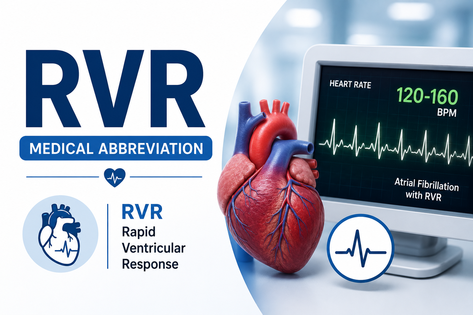
    RVR most often means rapid ventricular response in cardiology. Here is how to read the term on an ECG or hospital chart, why it commonly appears with AFib, and what determines whether it is an emergency.

    Medical context: RVR is a description of how fast the ventricles are responding to an atrial rhythm. It is not a stand-alone diagnosis. A fast or irregular pulse with chest pain, fainting, severe shortness of breath, confusion, or signs of shock needs urgent medical evaluation.

    The RVR medical abbreviation most commonly stands for rapid ventricular response, especially when it appears in an emergency department note, ECG report, cardiology visit, or hospital discharge summary. It is frequently used in the phrase “AFib with RVR,” meaning atrial fibrillation with a fast ventricular rate.

    The wording can sound more alarming than it is easy to interpret. Atrial fibrillation begins in the upper chambers of the heart, but the pulse you feel is created by the lower chambers, or ventricles. When too many irregular electrical impulses pass through the atrioventricular node and reach the ventricles, the ventricular rate can become rapid. That is the “RVR” part of the diagnosis.

    A rapid ventricular response can cause palpitations, shortness of breath, dizziness, fatigue, chest discomfort, or weakness. Some people have surprisingly few symptoms. The seriousness depends on more than the heart-rate number. Clinicians look at blood pressure, oxygenation, symptoms, heart function, the duration of the episode, other illnesses, and the exact rhythm on the ECG.

    Key Takeaways

    • RVR usually stands for rapid ventricular response, most commonly in the chart phrase “atrial fibrillation with RVR” or “AF with RVR.”
    • AFib and RVR are not the same thing. AFib describes the abnormal rhythm in the atria; RVR describes a fast response in the ventricles.
    • Clinicians often use RVR when the ventricular rate is above about 100 beats per minute, but there is no single cutoff that by itself determines severity or treatment.
    • An ECG is the key test for confirming AFib with RVR. Blood tests, echocardiography, monitoring, and other tests may be used to find triggers, complications, or underlying heart disease.
    • If AFib with RVR is causing hemodynamic instability, current American Heart Association guidance supports immediate synchronized electrical cardioversion.
    • Long-term AFib care addresses more than rate control. Stroke prevention, rhythm control when appropriate, blood pressure, sleep apnea, alcohol use, weight, exercise, and other risk factors can all matter.

    What Does RVR Stand For in Medical Terms?

    In cardiology, RVR stands for rapid ventricular response. The term describes a situation in which the ventricles are beating quickly because they are receiving frequent electrical signals from the atria.

    You may also hear clinicians say “rapid ventricular rate.” The intended meaning is usually similar: the lower chambers are contracting at an abnormally fast rate in response to an atrial arrhythmia. The abbreviation is most strongly associated with atrial fibrillation, although atrial flutter can also occur with a rapid ventricular response.

    RVR does not tell you why the atrial arrhythmia started, how long it has been present, whether a blood clot has formed, or whether the person needs a blood thinner. Those questions are handled separately during the evaluation.

    How Is AFib With RVR Different From AFib Alone?

    Atrial fibrillation is an irregular electrical rhythm in the atria. The atria quiver rather than contracting in a coordinated way. The AV node acts as a partial gatekeeper, preventing every atrial impulse from reaching the ventricles. How many impulses get through determines the ventricular rate.

    Term What it describes What you may see clinically
    AFib An irregular rhythm originating in the atria. The ventricular rate may be slow, normal, or fast.
    RVR A fast ventricular response to an atrial rhythm. A rapid pulse, often irregular when the underlying rhythm is AFib.
    AFib with RVR Atrial fibrillation plus a fast ventricular rate. Irregularly irregular rhythm with a rapid ventricular response on ECG.
    Atrial flutter with RVR Atrial flutter plus a rapid ventricular response. Atrial flutter waves with a fast ventricular rate, sometimes regular and sometimes variable.

    The distinction matters because the heart-rate problem and the rhythm problem are related but not identical. A clinician can slow the ventricular rate while the patient remains in AFib, or use a rhythm-control strategy to restore and maintain sinus rhythm when appropriate.

    What Heart Rate Counts as RVR?

    There is no universally binding number that turns atrial fibrillation into “AFib with RVR” in every clinical setting. Many clinicians use the term when the ventricular rate is greater than about 100 beats per minute at rest. Research studies and emergency protocols may use higher thresholds, such as 110, 120, or 150 beats per minute, depending on the question being studied.

    That is why a chart may say RVR at 115 beats per minute in one situation while another clinician may reserve the label for a faster rate. The more important issue is whether the rate is causing symptoms, poor blood pressure, inadequate organ perfusion, myocardial ischemia, worsening heart failure, or other signs of instability.

    A heart rate in the 130s may be tolerated by one stable patient for a short period but be dangerous for another person with severe heart failure, active coronary ischemia, dehydration, infection, or low blood pressure. Clinical context is more important than a single threshold.

    Why Can the Ventricular Rate Become So Fast?

    During AFib, the atria can generate hundreds of disorganized electrical impulses each minute. The AV node filters many of them, but if enough impulses pass through, the ventricles accelerate. Several factors can make that rapid response more likely or make an existing episode harder to control.

    • Infection, fever, or sepsis.
    • Dehydration or significant fluid shifts.
    • Hyperthyroidism or another thyroid abnormality.
    • Alcohol use, particularly binge drinking.
    • Stimulants or certain recreational drugs.
    • Missed doses or interruption of rate-control medication.
    • Acute heart failure or another major cardiac stressor.
    • Recent surgery or severe physical illness.
    • Poorly controlled high blood pressure.
    • Sleep-disordered breathing, including obstructive sleep apnea.
    • Electrolyte abnormalities or other metabolic problems.

    Sometimes AFib with RVR occurs without one obvious trigger. In those cases, the workup focuses on the patient’s broader AFib risk factors and cardiac history.

    What Does AFib With RVR Feel Like?

    Symptoms range from barely noticeable to severe. A rapid rate shortens the time the ventricles have to fill between beats, which can reduce effective cardiac output. The loss of coordinated atrial contraction from AFib can add to that effect.

    • A racing, pounding, fluttering, or irregular heartbeat.
    • Shortness of breath, especially with activity.
    • Lightheadedness or dizziness.
    • Weakness or unusual fatigue.
    • Reduced exercise tolerance.
    • Chest pressure or discomfort.
    • Sweating or a feeling of anxiety.
    • Near-fainting or fainting in more severe episodes.

    AFib can also be asymptomatic. A person may first learn about it after a routine pulse check, ECG, wearable alert, or examination for another condition.

    For a patient-friendly overview of AFib symptoms and complications, see the National Heart, Lung, and Blood Institute atrial fibrillation page.

    How Do Doctors Confirm RVR?

    The central test is an electrocardiogram, or ECG. A 12-lead ECG can show the atrial rhythm and the ventricular rate at the same time. In AFib, clinicians typically look for an irregularly irregular ventricular rhythm without consistent P waves.

    If the episode comes and goes, a normal ECG in the clinic does not necessarily rule it out. Depending on symptom frequency, a clinician may use longer monitoring.

    • A Holter monitor for continuous short-term recording.
    • A patch monitor for several days or longer.
    • An event monitor for intermittent symptoms.
    • An implantable loop recorder in selected patients with infrequent but concerning episodes.
    • A consumer wearable or smartwatch tracing as a clue that still needs clinical confirmation.

    What Other Tests May Be Ordered?

    RVR is a rhythm description, so the next step is often to search for the trigger and assess the heart. Testing is individualized, but may include:

    • Blood count to look for anemia or infection.
    • Electrolytes and kidney function, which can affect rhythm and medication choices.
    • Thyroid testing when hyperthyroidism or another thyroid disorder is possible.
    • Cardiac biomarkers when symptoms suggest myocardial injury or acute coronary syndrome.
    • Echocardiography to assess heart size, valves, ejection fraction, and structural disease.
    • Chest imaging when lung disease, pulmonary congestion, or another cardiopulmonary problem is suspected.

    When Is AFib With RVR an Emergency?

    The most important distinction in acute care is whether the patient is hemodynamically stable. Current 2025 American Heart Association advanced life support guidance recommends immediate electrical cardioversion when hemodynamic instability is attributable to atrial fibrillation or atrial flutter with rapid ventricular rates.

    Signs that can point to instability include:

    • Very low blood pressure or signs of shock.
    • New or worsening confusion or altered mental status.
    • Ongoing ischemic chest discomfort.
    • Acute heart failure or severe breathing difficulty.
    • Fainting or evidence that organs are not being adequately perfused.

    A fast pulse alone does not prove that the rhythm is causing instability. For example, sepsis can produce both low blood pressure and a rapid heart rate. Emergency clinicians determine whether the arrhythmia is the cause, a contributor, or a consequence of another critical illness.

    The current U.S. emergency-care recommendations are summarized in the American Heart Association 2025 Adult Advanced Life Support guideline.

    How Is AFib With RVR Treated in the Emergency Department?

    Treatment starts with stability. If the patient is unstable because of the arrhythmia, synchronized electrical cardioversion is the urgent treatment. If the patient is stable, clinicians usually focus first on slowing the ventricular rate, correcting triggers, and deciding whether a rhythm-control strategy is appropriate.

    Rate Control

    For hemodynamically stable AFib or atrial flutter with RVR and no preexcitation, commonly used IV rate-control drugs include beta blockers and nondihydropyridine calcium channel blockers. Examples include metoprolol or esmolol in the beta-blocker group and diltiazem or verapamil in the calcium channel blocker group.

    Drug choice is not interchangeable in every patient. Diltiazem and verapamil can worsen hemodynamics in people with left ventricular systolic dysfunction or decompensated heart failure. Beta blockers also require caution in decompensated states. In critically ill patients who cannot use those options, IV amiodarone may sometimes be used for rate control. Digoxin has a slower onset and is less commonly relied on for immediate control in the emergency setting.

    Rhythm Control

    Rhythm control aims to restore sinus rhythm rather than simply slow the ventricular rate. This can involve synchronized cardioversion, antiarrhythmic medication, or a longer-term strategy such as catheter ablation. The choice depends on symptoms, duration of AFib, prior episodes, heart structure and function, comorbidities, treatment history, and patient preference.

    Treating the Trigger

    A rate-control medication may not work well if the underlying driver is still active. Treatment may also involve IV fluids for appropriate patients with dehydration, correction of electrolyte abnormalities, treatment of infection, management of hyperthyroidism, care for acute heart failure, or stopping a provoking substance or medication when medically appropriate.

    Why Does Preexcitation Change the Treatment?

    A small but important group of patients has AFib with an accessory electrical pathway, such as Wolff-Parkinson-White syndrome. This is called preexcited AFib. In that setting, drugs that slow conduction through the AV node can unintentionally encourage more impulses to travel down the accessory pathway, producing an extremely rapid ventricular rate and potentially ventricular fibrillation.

    The 2025 AHA advanced life support guidance warns against AV nodal blocking drugs such as digoxin, nondihydropyridine calcium channel blockers, beta blockers, and IV amiodarone in preexcited AFib or flutter. This is one reason an ECG diagnosis should guide treatment rather than trying to manage a fast irregular pulse at home with leftover medication.

    Does RVR Increase Stroke Risk?

    The stroke risk comes primarily from atrial fibrillation itself rather than from the RVR label. During AFib, ineffective atrial contraction can allow blood to pool, particularly in the left atrial appendage, where a clot can form and later travel to the brain.

    Whether someone needs anticoagulation is based on estimated thromboembolic risk and the individual clinical situation. It is not decided simply by whether the ventricular rate was 90, 120, or 160 beats per minute.

    Current U.S. AFib guidance uses validated stroke-risk assessment and generally favors direct oral anticoagulants over warfarin for eligible patients, with important exceptions such as mechanical heart valves and certain mitral stenosis scenarios. Aspirin is not considered an equivalent substitute for anticoagulation in patients who otherwise qualify for it.

    The broader U.S. approach to AFib stroke prevention and long-term management is outlined in the ACC summary of the 2023 ACC/AHA/ACCP/HRS atrial fibrillation guideline.

    Can RVR Damage the Heart?

    A persistently rapid ventricular rate can place substantial stress on the heart. If the rate remains uncontrolled for long enough, some patients can develop or worsen heart failure. Sustained tachycardia can also contribute to tachycardia-induced cardiomyopathy, a potentially reversible weakening of the heart muscle caused by prolonged rapid rhythm.

    The risk depends on how fast the heart is beating, how long the abnormal rate lasts, and the person’s underlying heart function. Someone with a structurally normal heart may tolerate a short episode better than someone with severe valve disease, cardiomyopathy, coronary disease, or preexisting heart failure.

    This is why recurrent RVR episodes deserve follow-up even when they stop on their own. The long-term goal is not simply to survive each fast-rate episode but to reduce symptom burden, prevent complications, and address the AFib itself.

    What Is the Difference Between Rate Control and Rhythm Control?

    Strategy Main goal Common examples
    Rate control Keep the ventricular rate in a safer, more tolerable range while AFib may continue. Beta blockers, diltiazem, verapamil, digoxin in selected patients.
    Rhythm control Restore and maintain sinus rhythm when appropriate. Cardioversion, antiarrhythmic drugs, catheter ablation.
    Stroke prevention Reduce the chance of an AFib-related clot reaching the brain. Anticoagulation when indicated by stroke risk and clinical factors.
    Risk-factor treatment Reduce AFib burden and progression over time. Blood pressure control, weight management, exercise, sleep apnea evaluation, limiting alcohol, tobacco cessation.

    Modern AFib treatment increasingly combines these strategies rather than treating them as mutually exclusive. A patient may need rate control today, rhythm control later, anticoagulation for stroke prevention, and risk-factor treatment throughout the course of care.

    Can a Smartwatch Detect AFib With RVR?

    Wearable devices can be useful for detecting an unexpectedly fast or irregular pulse, and some devices can record a single-lead ECG that may identify possible atrial fibrillation. They can be particularly helpful when symptoms are intermittent.

    A wearable alert is not the same as a clinical ECG diagnosis. Motion artifact, premature beats, sinus tachycardia, atrial flutter, and other rhythms can create confusing readings. If a device repeatedly shows a fast irregular rhythm, especially with symptoms, save the tracing if possible and contact a clinician.

    If the alert occurs with chest pain, fainting, severe shortness of breath, new neurological symptoms, or signs of shock, the appropriate response is urgent medical care rather than repeatedly checking the watch.

    What Can Trigger Repeat RVR Episodes?

    Preventing recurrence depends partly on what is driving AFib. Some triggers are modifiable, while others reflect underlying heart disease or aging. A practical long-term review may include:

    • Taking prescribed rate-control, rhythm-control, and anticoagulant medicines consistently.
    • Limiting or eliminating alcohol if it triggers AFib.
    • Managing high blood pressure and diabetes.
    • Treating sleep apnea when present.
    • Working toward a healthy weight if overweight or obesity contributes to AFib risk.
    • Maintaining regular exercise at a level recommended by the treating clinician.
    • Avoiding tobacco and discussing stimulant exposure with a clinician.
    • Following up after AFib discovered during infection, surgery, or another acute illness because recurrence can still occur later.

    When Should You Call 911?

    For someone with a known or suspected rapid irregular heart rhythm, call 911 in the United States when there are signs of a potentially life-threatening problem, including severe or persistent chest pain, fainting, severe trouble breathing, new confusion, symptoms of stroke, or signs of shock. Do not drive yourself if you are severely symptomatic or feel as though you may pass out.

    If the pulse is fast but you are otherwise stable, contact your clinician for individualized instructions. People with known AFib may have a specific action plan for recurrent episodes. Do not take extra doses of beta blockers, calcium channel blockers, antiarrhythmics, or blood thinners unless your treating clinician has specifically instructed you to do so.

    Questions People Ask About the RVR Abbreviation

    Does RVR always mean AFib?

    No. RVR describes a rapid ventricular response and can appear with other atrial rhythms, especially atrial flutter. In everyday hospital documentation, however, “RVR” is most commonly seen with AFib.

    Is AFib with RVR worse than regular AFib?

    It can be more symptomatic and can place more immediate stress on the heart because the ventricular rate is fast. Severity still depends on blood pressure, symptoms, heart function, duration, and other medical conditions.

    Can AFib with RVR go away on its own?

    Yes. Some paroxysmal AFib episodes terminate spontaneously, and the ventricular rate can also slow. A self-terminating episode still deserves medical review when it is new, recurrent, prolonged, or symptomatic because stroke risk and the need for long-term treatment are separate questions.

    Is a heart rate over 100 automatically RVR?

    Not automatically. Many clinicians use a rate above about 100 beats per minute as a practical RVR threshold in AFib, but the term is not defined by one universal cutoff. The ECG rhythm and clinical condition matter more than a single number.

    Can anxiety cause RVR?

    Anxiety can increase heart rate through sympathetic activation and may make palpitations more noticeable. It does not by itself diagnose AFib with RVR. A fast irregular rhythm should be confirmed with ECG monitoring rather than assumed to be anxiety.

    Can dehydration cause AFib with RVR?

    Dehydration can contribute to a fast heart rate and may be one trigger in a susceptible person, particularly when combined with illness, heat exposure, alcohol, or electrolyte changes. Clinicians still look for other causes and treat the actual rhythm.

    What does “controlled ventricular response” mean?

    It usually means AFib is still present but the ventricular rate is no longer excessively fast. The exact target is individualized based on symptoms, heart function, activity level, and the treatment strategy.

    Can you have RVR without feeling it?

    Yes. Some people have little or no awareness of a rapid irregular rhythm. That is one reason AFib may be discovered on a routine examination, ECG, or wearable device rather than because of obvious palpitations.

    The Bottom Line

    In most cardiology notes, the RVR medical abbreviation means rapid ventricular response. It most often appears as “AFib with RVR,” which tells you that atrial fibrillation is present and the ventricles are responding at a fast rate.

    The abbreviation is useful, but it is only one piece of the clinical picture. A rate above about 100 beats per minute is commonly described as rapid, yet treatment is driven by the ECG, symptoms, blood pressure, heart function, underlying cause, duration of AFib, and stroke risk. Unstable AFib with RVR may require immediate synchronized cardioversion, while stable episodes are often managed with carefully selected rate-control or rhythm-control treatment plus attention to triggers and long-term AFib care.

    If RVR appears on your ECG or discharge paperwork, the most useful follow-up questions are not just “How fast was my heart?” Ask what rhythm caused it, whether your heart structure and function are normal, what your stroke risk is, what should trigger emergency care, and what plan can reduce another episode.

    Medical Review Note

    This article is for general education and is not a substitute for diagnosis or individualized treatment. The content is written for a U.S. audience and reflects information available in 2026, including the 2023 ACC/AHA/ACCP/HRS atrial fibrillation guideline and the American Heart Association 2025 Adult Advanced Life Support recommendations.

  • Liver Disease and Pale Stool: What Clay, Gray, and Light-Colored Stool Can Look Like

    Liver Disease and Pale Stool: What Clay, Gray, and Light-Colored Stool Can Look Like

    People searching for information about liver disease and pale stool are often concerned that an unusually light bowel movement could signal reduced bile flow. Pale stool can be associated with liver or bile duct problems, but color alone cannot diagnose liver disease. This 2026 U.S.-focused guide explains the visual differences between pale, clay-colored, yellow, greasy, and normal brown stool, plus the symptoms that should prompt medical care.

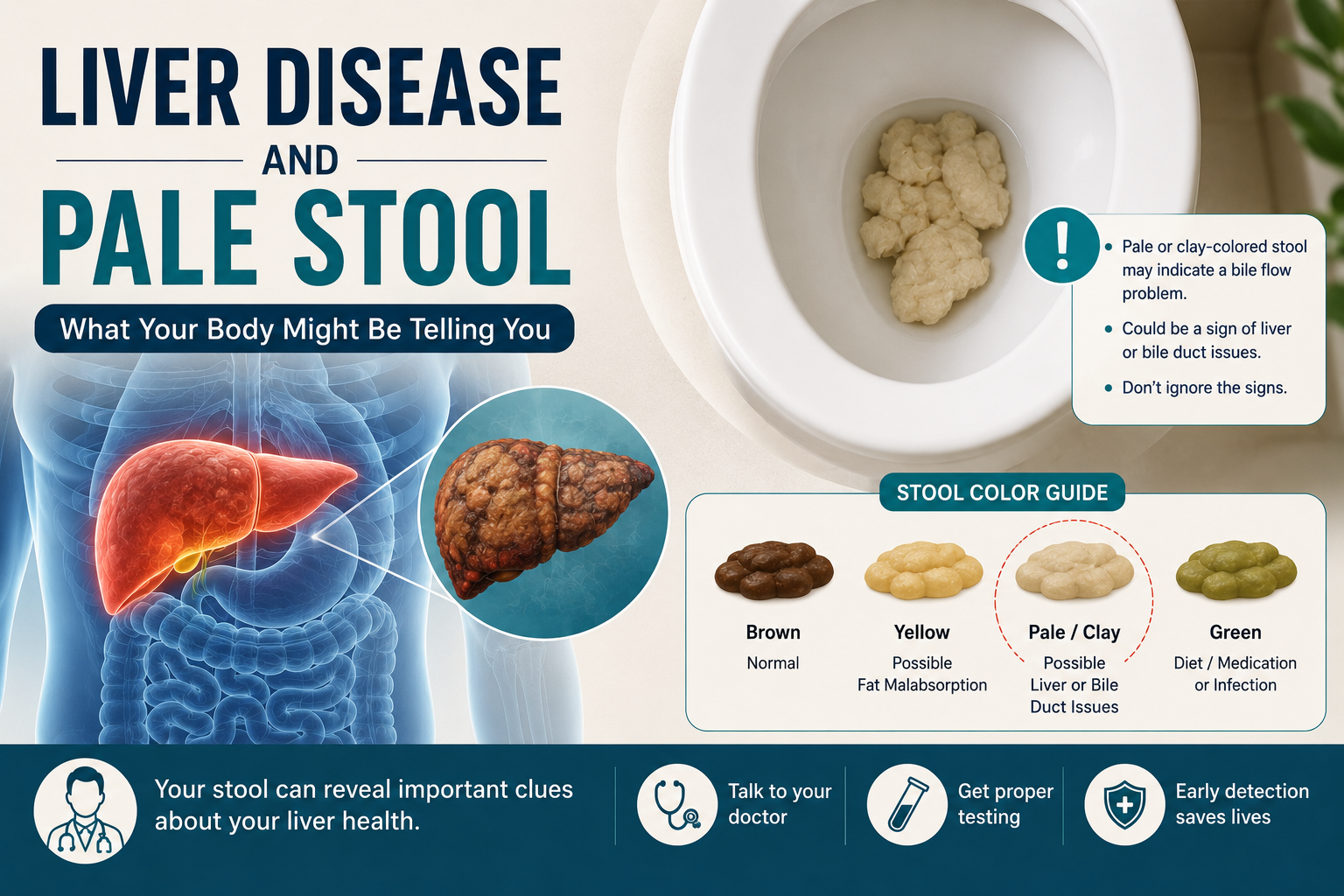
    People searching for liver disease pale stool pictures are often trying to decide whether an unusual bowel movement is simply a temporary color change or a sign that bile flow has been reduced. Normal stool is usually some shade of brown because bile pigments are changed as they pass through the digestive tract. When little or no bile reaches the intestine, stool may become unusually light, gray, putty-colored, or nearly white.

    A single lighter-than-usual bowel movement is not enough to diagnose a liver, gallbladder, bile duct, or pancreatic problem. Persistently pale or clay-colored stools deserve more attention, especially when they occur with dark urine, yellow skin or eyes, itching, fever, or abdominal pain. MedlinePlus explains that pale, clay, or putty-colored stool can occur with problems in the biliary system.

    Key Takeaways

    • Truly pale stool is usually described as clay-colored, putty-colored, gray, very light beige, or almost white rather than simply light brown.
    • Reduced bile reaching the intestine is an important reason stool can lose its normal brown color.
    • Liver disease is only one possible cause. Gallstones, bile duct obstruction, pancreatic disease, certain medications, and medical procedures can also change stool color.
    • Yellow stool is not the same as clay-colored stool. Yellow stool may occur with diet changes, diarrhea, rapid intestinal transit, infections, or fat malabsorption.
    • Pale stool together with jaundice or dark urine is more concerning than an isolated light-colored bowel movement.
    • Persistent white, gray, or clay-colored stool should be discussed with a health care professional rather than diagnosed from pictures alone.

    What does pale stool from reduced bile look like?

    There is no single exact shade that proves bile flow is abnormal. Lighting, toilet water, diet, camera settings, and the natural variation of stool can all affect what a picture looks like. Instead of matching one online image, focus on whether the stool has clearly lost its usual brown pigment and whether the change keeps happening.

    Appearance Typical description What it may suggest How concerning is it?
    Normal brown Tan-brown to dark brown Normal bile pigment in the digestive tract Usually normal if consistent for you
    Light brown or beige Brown is still clearly visible but lighter than usual Diet, temporary digestive change, or normal variation Often less concerning if brief and no other symptoms
    Clay or putty-colored Very pale beige, gray-beige, or putty-like Reduced bile entering the intestine can be one cause Needs evaluation if persistent
    Gray or almost white Little visible brown pigment Bile duct, liver, gallbladder, or pancreatic problems may need to be ruled out More concerning, especially with jaundice or dark urine
    Yellow and greasy Yellow, oily, floating, difficult to flush, sometimes foul-smelling Fat malabsorption or rapid transit may be involved Needs evaluation if recurrent or associated with weight loss
    Bright yellow loose stool Yellow diarrhea rather than pale formed stool Diet, infection, rapid intestinal transit, or other GI causes Context and duration matter

    Why does liver or bile duct disease make stool pale?

    The liver produces bile, a fluid that helps the body digest fats. Bile travels through small ducts into larger bile ducts and eventually reaches the small intestine. Pigments from bile contribute to the brown color of stool.

    If bile production is severely impaired or bile cannot flow normally through the ducts, less pigment reaches the intestine. The stool can therefore lose its brown color and become light, gray, clay-colored, or nearly white. This is sometimes called acholic stool.

    The important point is that pale stool reflects a possible bile-flow problem, not one particular liver diagnosis. A blockage outside the liver can produce a similar color change to a disorder affecting bile formation inside the liver.

    Which liver problems can be associated with pale stool?

    Several liver conditions can interfere with bile production or bile flow, although pale stool is not present in every person with liver disease.

    • Hepatitis: Inflammation of the liver from viral infection, medications, toxins, autoimmune disease, or other causes may sometimes occur with jaundice, dark urine, and pale or clay-colored stool.
    • Cirrhosis: Advanced scarring can disrupt normal liver function and, in some cases, contribute to jaundice or bile-flow abnormalities.
    • Cholestatic liver disease: Conditions that interfere with the formation or movement of bile can lead to itching, jaundice, dark urine, and pale stool.
    • Liver or bile duct tumors: A mass can sometimes narrow or block bile drainage, although many other conditions can cause the same symptom pattern.

    NIDDK notes that people with cirrhosis may develop jaundice and other signs of impaired liver function as disease progresses. Read the NIDDK overview of cirrhosis symptoms and causes.

    Can gallstones cause pale or clay-colored stool?

    Yes. Gallstones can block a bile duct and prevent bile from moving normally into the intestine. When a duct is blocked, a person may develop light-colored stool along with pain in the upper right or upper middle abdomen, nausea, vomiting, fever or chills, jaundice, or dark urine.

    A blocked bile duct can lead to serious complications and should not be ignored. NIDDK lists light-colored stools among warning symptoms that can occur when gallstones cause complications.

    Can pancreatic problems make stool look pale?

    They can. The pancreas sits near the bile duct and supplies digestive enzymes to the small intestine. Pancreatic inflammation, a pancreatic mass, or another condition that interferes with bile drainage can sometimes contribute to pale stool.

    Pancreatic disorders can also cause a different pattern called steatorrhea. This stool is usually greasy, oily, bulky, foul-smelling, difficult to flush, or prone to floating because excess fat remains in the stool. It may look yellowish or light, but greasy yellow stool is not automatically the same as bile-deprived clay-colored stool.

    Is yellow stool a sign of liver disease?

    Sometimes, but yellow stool has many non-liver causes. A one-time yellow bowel movement can follow certain foods or a bout of diarrhea. When stool passes through the intestine quickly, bile pigments may not undergo the same color changes that normally produce brown stool.

    Yellow, greasy, floating stool can also occur with fat malabsorption. Possible causes include pancreatic enzyme problems, celiac disease, and other digestive disorders. By contrast, the classic bile-flow concern is stool that becomes unusually pale, gray, clay-colored, or white.

    What other things can make stool look unusually pale?

    • Medications and contrast materials: Some medicines or substances used for imaging tests can temporarily lighten stool.
    • Recent diarrhea: Fast intestinal transit can temporarily change stool color and consistency.
    • Dietary changes: Very light foods or a sudden change in diet can alter appearance, although diet alone usually does not produce repeated chalky-white stool.
    • Lighting and toilet water: Bathroom lighting and dilution in water can make tan or yellow stool look paler in photographs than it does in person.

    What symptoms make pale stool more concerning?

    Stool color becomes more medically important when other symptoms point toward impaired bile flow, liver inflammation, infection, or obstruction.

    • yellowing of the skin or whites of the eyes
    • dark brown or tea-colored urine
    • persistent itching without an obvious skin cause
    • pain in the upper right or upper middle abdomen
    • fever or chills
    • nausea or vomiting that persists
    • unexplained weight loss or loss of appetite
    • abdominal swelling
    • new confusion or unusual drowsiness
    • repeated gray, white, or clay-colored bowel movements

    Pale stool with jaundice and dark urine is a particularly important combination because it can occur when bilirubin is building up in the blood while too little bile pigment is reaching the intestine.

    When should you call a doctor about pale stool?

    Contact a health care professional if stool remains clay-colored, gray, white, or unusually pale for more than a short period, or if the change keeps returning. You should also seek medical care sooner when pale stool occurs with jaundice, dark urine, significant abdominal pain, fever, vomiting, or unexplained weight loss.

    Severe abdominal pain, high fever, confusion, fainting, repeated vomiting, or signs of serious illness warrant urgent evaluation. A picture of the stool may be useful to show a clinician, but it does not replace examination and testing.

    How do doctors evaluate persistent pale stool?

    Evaluation depends on symptoms and medical history. A clinician may ask how long the color change has lasted, whether the stool is greasy or floating, what medications and supplements you take, alcohol use, recent travel or infection risk, and whether you have jaundice, abdominal pain, or weight loss.

    • Blood tests: Liver enzymes, bilirubin, alkaline phosphatase, and other laboratory tests can help identify patterns of liver injury or impaired bile flow.
    • Ultrasound: Abdominal ultrasound can look for gallstones, bile duct dilation, and some liver or gallbladder abnormalities.
    • CT or MRI: Cross-sectional imaging may be ordered when a blockage, pancreatic problem, or other structural cause is suspected.
    • Additional testing: Depending on the findings, doctors may use specialized bile-duct imaging, viral hepatitis tests, pancreatic tests, or other studies.

    Can you diagnose liver disease from a pale stool picture?

    No. Pictures can help illustrate color ranges, but they cannot determine why stool is pale. Several unrelated conditions can produce similar-looking bowel movements, and normal lighting differences can make online comparisons misleading.

    A better approach is to note the color over several bowel movements, whether the stool is formed or loose, whether it floats or appears greasy, and whether symptoms such as jaundice, dark urine, pain, fever, itching, or weight loss are present. That information is much more useful to a clinician than trying to match one photograph.

    Common Questions About Liver Disease and Pale Stool

    What color is stool with liver disease?

    There is no single liver-disease stool color. Some conditions that reduce bile reaching the intestine can produce pale, gray, clay-colored, or nearly white stool. Many people with liver disease continue to have brown stool.

    Is light brown stool the same as clay-colored stool?

    Usually not. Light brown stool still contains obvious brown pigment. Clay-colored stool is noticeably washed out, beige-gray, putty-like, or nearly white.

    Can fatty liver disease cause pale stool?

    Fatty liver disease does not automatically cause pale stool. If a person with known liver disease develops persistent clay-colored stool, jaundice, or dark urine, the change should be medically evaluated rather than assumed to come from fatty liver alone.

    Does pale stool always mean a blocked bile duct?

    No. Bile duct blockage is an important possibility, but medications, liver disease, pancreatic disease, and other conditions can also contribute. Testing is needed to identify the cause.

    What does greasy yellow stool mean?

    Greasy, oily, floating, or difficult-to-flush yellow stool may suggest excess fat in the stool, called steatorrhea. This can occur with pancreatic or intestinal malabsorption problems and is different from classic clay-colored stool.

    Should I save a picture of unusual stool for my doctor?

    A clear photo can help document an intermittent color change, especially if the stool looks different by the time of the appointment. It should be treated as supporting information, not as a diagnostic test.

    What is the bottom line on pale stool and liver disease?

    Liver disease can be associated with pale stool, but the more direct issue is usually reduced bile reaching the intestine. That can happen because of liver disease, gallstones, bile duct obstruction, or pancreatic and other biliary problems.

    A one-time light stool may be harmless, especially if it is still clearly brown. Repeated gray, white, clay-colored, or putty-colored stool deserves attention, particularly when it appears with jaundice, dark urine, itching, abdominal pain, fever, or unexplained weight loss. Online pictures can help with visual comparison, but persistent color changes are best evaluated with medical history, blood tests, and imaging when needed.

    Medical Note

    This article is for general educational purposes and is not a substitute for medical advice, diagnosis, or treatment. Persistent pale stool or stool changes accompanied by jaundice, dark urine, fever, significant abdominal pain, or unexplained weight loss should be discussed with a qualified health care professional.

    Editorial Sources

    This 2026 update was informed by current U.S. information from MedlinePlus and the National Institute of Diabetes and Digestive and Kidney Diseases. The HealthCentral reference was used for search intent and topic coverage only.